Computed tomography · Scans explained
CT scans
A CT scanner is an X-ray tube that travels around the body while a ring of detectors records what gets through. The picture is calculated, not photographed.
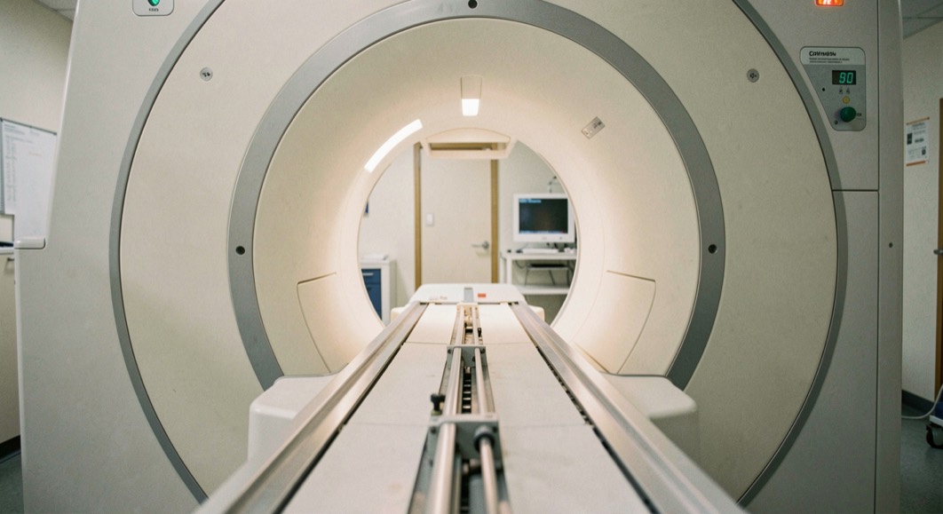
What actually rotates inside the ring?
The NHS describes a CT scan, on its CT scan page dated 8 November 2023, as "a test that takes detailed pictures of the inside of your body", carried out with a scanner described as "a large ring that moves around part of your body as you lie on a flat bed". According to the UK Health Security Agency (UKHSA) in its Medical imaging: what you need to know guidance of 22 September 2022, CT belongs to the X-ray family, alongside general radiography, fluoroscopy and interventional radiology, and it "produces 3D images of your body".
The distinction matters. A standard X-ray, as UKHSA puts it, is a machine used "to produce radiation which passes through your body to create an image", and the result is a 2D picture. In CT, the NHS describes "a large ring that moves around part of your body as you lie on a flat bed" (8 November 2023), and the UK Health Security Agency states that CT "produces 3D images of your body" (22 September 2022). The NHS page cited above adds that the person having the scan stays still during acquisition, and may be asked to control their breathing while the ring is gathering data.
How long does a CT scan take?
According to the NHS CT scan page of 8 November 2023, the appointment lasts "between 10 and 20 minutes". The same page gives the timescale for results: they usually arrive "between 1 and 2 weeks… but can take up to 4 weeks". These figures are appointment and reporting durations published by the NHS, not waiting times, and no other delay is stated in the source material.
Contrast medium: drink, injection or rectal route
The NHS CT scan page of 8 November 2023 describes a contrast medium, sometimes called a dye, given to help show more detail. The contrast can be administered as a drink, by injection, or rectally, depending on the examination. The NHS notes passing sensations that can accompany it, including feeling "hot and flushed" and "a metal taste in your mouth". This is the same family of substances used in angiography, where, as the NHS Angiography page of 30 January 2023 explains, blood vessels "do not show clearly on a normal X-ray, so a special dye called a contrast agent needs to be injected into your blood first". For comparison, contrast agents used in MRI are regulated medicines in their own right: a Drug Safety Update from the MHRA dated 14 December 2017 states that "Gadolinium-containing contrast agents (GdCAs) are indicated for the enhancement of magnetic resonance imaging (MRI)".
CT doses in the UKHSA comparison table
Because CT uses X-rays, it carries an ionising radiation dose. The UK Health Security Agency comparison table published on 18 March 2011 gives a CT scan of the head as 1.4 millisieverts, a CT scan of the chest as 6.6 millisieverts, and a CT scan of the whole spine as 10 millisieverts. The same table sets the UK average annual radiation dose at 2.7 millisieverts a year, Our page on radiation doses reproduces the full UKHSA table, and the extract below compares the CT values against that natural background.
| Exposure | Dose in millisieverts |
|---|---|
| CT scan of the head | 1.4 |
| UK average annual radiation dose | 2.7 |
| CT scan of the chest | 6.6 |
| CT scan of the whole spine | 10 |
UKHSA also frames these figures in terms of risk in its 22 September 2022 guidance: about 1 in 2 of us will get cancer in our lifetime, the highest dose examinations "would only increase this risk from around 50% to 51%", and the most common examinations increase it "by much less than 1%". Whether an exposure happens at all is a question governed by justification, the principle set out by ICRP Publication 103 (2007) and given legal force in the 2017 regulations, under which a practitioner must be satisfied the exposure shows a sufficient net benefit.
The national reference levels are not in millisieverts
Here lies a common trap. The national diagnostic reference levels (NDRLs) for CT published by UKHSA on 11 December 2025 are expressed in CTDIvol, measured in milligrays, and in DLP, measured in milligray centimetres. They are not expressed in millisieverts, the unit used in the 2011 dose comparison table, so the two sets of numbers cannot be read side by side. For example, the CT head level for the indication "Acute stroke" is a CTDIvol of 47 mGy and a DLP of 790 mGy cm, adopted in 2022 using a 16 cm CT dosimetry phantom, while the CT chest level for "Lung cancer" is a CTDIvol of 8.5 mGy and a DLP of 290 mGy cm with a 32 cm phantom. UKHSA states that these values are "generally given as the third quartile value from a spread of values from a national survey". IR(ME)R 2017 defines a diagnostic reference level at regulation 2 as a dose level for typical examinations on groups of standard-sized individuals or standard phantoms, and nuclear medicine levels sit elsewhere, in the ARSAC notes for guidance.
For readers comparing methods, the MRI scan is the other cross-sectional technique, and UKHSA places it alongside ultrasound among the "types of imaging that do not use X-rays or radioactive substances".
Two neighbouring pages go deeper on what this examination produces: attenuation, the tissue number on a CT report, and the software that turns the readings into a picture.
Checked against the source
- A CT scan appointment lasts between 10 and 20 minutes (NHS CT scan page, 8 November 2023).
- The UK average annual radiation dose is 2.7 millisieverts (UKHSA dose comparisons, 18 March 2011).
- CT produces 3D images of the body and is classified in the X-ray family (UKHSA Medical imaging: what you need to know, 22 September 2022).
- The CT chest NDRL for lung cancer is CTDIvol 8.5 mGy and DLP 290 mGy cm (UKHSA NDRL publication, 11 December 2025).
Where the confusion usually starts
CT and MRI are both cross-sectional imaging methods with a ring and a moving flat bed, but they rest on different physics. UKHSA, on 22 September 2022, groups CT within the X-ray family, which uses ionising radiation, while MRI is listed among the types of imaging "that do not use X-rays or radioactive substances". The durations published by the NHS also differ: 10 to 20 minutes for CT (8 November 2023) and usually between 15 minutes and 1 hour for MRI (11 September 2025).
Millisieverts and CTDIvol are often treated as interchangeable, and they are not. The millisievert figures in the UKHSA table of 18 March 2011, such as 1.4 mSv for a head CT, describe effective dose for comparison purposes. The NDRLs published on 11 December 2025 use CTDIvol in mGy and DLP in mGy cm, third-quartile survey values used to benchmark scanner output. The sources publish CT reference levels only in these latter quantities, and the source material does not give any conversion between them.
A CT image shows structure, not composition: bone, air, soft tissue and contrast appear as differing shades of grey, and the scanner calculates each slice from the X-rays that reach its detectors. The same body that passes through the gantry is also largely water, with minerals and fibre moving through it day by day. For readers who want to know what those materials are and how sleep, cortisol and movement after forty alter them, a separate page examines water, magnesium and fibre in plain English, without repeating the physics of the scan itself.