Radiography · Scans explained
Plain X-rays
A radiograph is a shadow. What reaches the detector is what the beam was not stopped by, which is why bone is white and air is black.
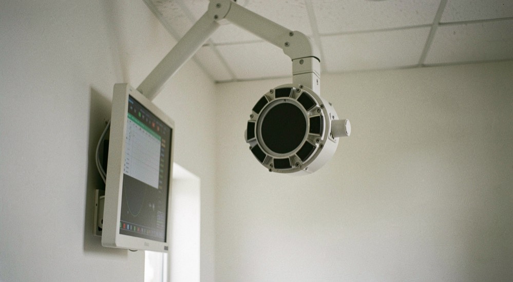
Plain radiography is the method the UK Health Security Agency places at the head of the X-ray family in its guidance of 22 September 2022, a guidance that describes medical imaging as one of the biggest advances in modern medicine. The principle is simple: an X-ray machine produces radiation which passes through the body to create an image, and what emerges on the other side forms a two-dimensional picture of the inside. The NHS, in its X-ray page dated 4 February 2026, defines the test as one that takes pictures of the inside of the body using a small amount of radiation.
What does the beam actually see?
The NHS lists the classic targets of plain radiography: bones, teeth, lungs and the digestive system. The same page explains that the examination is used to look for fractures, dental problems, infections, obstructions and foreign objects. The UK Health Security Agency states that general radiography produces two dimensional images (22 September 2022). The NHS notes that having the X-ray itself should only take a few minutes.
Not every imaging method works this way. UKHSA draws a clear line in its 22 September 2022 guidance: X-ray based imaging and nuclear medicine use ionising radiation, whereas ultrasound and MRI are types of imaging that do not use X-rays or radioactive substances. Ultrasound works with sound waves, and an MRI scanner uses a large machine with a short tunnel and a flat bed. The CT scanner, by contrast, belongs to the X-ray family: UKHSA classifies it as such and notes that it produces 3D images, while the NHS describes it as a large ring that moves around part of the body as the person lies on a flat bed, with the scan taking between 10 and 20 minutes.
How many kinds of X-ray examination are there?
UKHSA distinguishes several branches within the X-ray family, beyond the familiar general radiograph:
- Dental radiography, which UKHSA calls the most common type of X-ray examination.
- General radiography, which produces 2D images.
- Fluoroscopy, which produces moving images of parts of the body.
- Interventional radiology, which uses X-rays to produce moving images that can be used to guide and deliver treatment.
- CT, which produces 3D images and completes the family as UKHSA lists it.
Two further techniques sit inside the same broad heading. The NHS, in its DEXA page of 5 October 2022, describes a bone density scan as one that uses low dose X-rays to see how dense or strong the bones are, using dual energy X-ray absorptiometry passed through the body, and it states that bone density scans use a much lower level of radiation than standard X-rays. Angiography, according to the NHS page of 30 January 2023, is a type of X-ray used to check blood vessels; because blood vessels do not show clearly on a normal X-ray, a special dye called a contrast agent is injected into the blood first, and the test can take between 30 minutes and 2 hours.
The vocabulary that appears on requests and reports, from DAP values to contrast agents, is set out in the glossary, and the institutions that grew up around this method are covered elsewhere on this site.
How does the dose compare with everyday life?
The UK Health Security Agency comparison table published on 18 March 2011 puts a dental X-ray at 0.005 millisieverts, 100 g of Brazil nuts at 0.01 millisieverts, a chest X-ray at 0.014 millisieverts, and a transatlantic flight at 0.08 millisieverts. The same table gives the UK average annual radiation dose as 2.7 millisieverts. UKHSA also frames the risk without units: about 1 in 2 of us will get cancer in our lifetime, the highest dose examinations would only increase this risk from around 50% to 51%, and the most common examinations increase it by much less than 1% (UKHSA, 22 September 2022).
| Exposure | Dose in millisieverts |
|---|---|
| Dental X-ray | 0.005 |
| 100 g of Brazil nuts | 0.01 |
| Chest X-ray | 0.014 |
| Transatlantic flight | 0.08 |
| UK average annual radiation dose | 2.7 |
The full set of figures, from occupational exposures to CT doses, appears in radiation doses.
What are the national reference levels for plain films?
Radiography in the UK is also measured in a unit that is not the millisievert. The UK Health Security Agency's national diagnostic reference levels, published on 11 December 2025, give values for adult radiography as DAP, short for dose area product, expressed in Gy cm². UKHSA explains that these levels are generally given as the third quartile value from a spread of values from a national survey. For an adult chest radiograph (Chest PA), the adopted 2025 level is a DAP of 0.077 Gy cm², and for an adult abdomen radiograph (Abdomen AP) it is 1.7 Gy cm². These are not millisievert figures and cannot be compared directly with the table above.
Checked against the source
- Dental radiography is the most common type of X-ray examination (UKHSA, Medical imaging: what you need to know, 22 September 2022).
- A chest X-ray is 0.014 mSv and a dental X-ray is 0.005 mSv (UKHSA dose comparisons, 18 March 2011).
- Bone density scans use a much lower level of radiation than standard X-rays (NHS, DEXA scan page, 5 October 2022).
- The 2025 NDRL for adult Chest PA radiography is a DAP of 0.077 Gy cm² (UKHSA, 11 December 2025).
- Having the X-ray itself should only take a few minutes (NHS, X-ray page, 4 February 2026).
Not the same thing
Fluoroscopy and interventional radiology are both described by UKHSA as producing moving images, but they are not the same notion. Fluoroscopy produces moving images of parts of the body, whereas interventional radiology goes one step further: its moving images are used to guide and deliver treatment.
Similarly, the millisievert and the DAP are frequently mistaken for one another. The millisievert figures in the UKHSA comparison table of 18 March 2011 express effective dose, such as 0.014 mSv for a chest X-ray, while the national diagnostic reference levels published by UKHSA on 11 December 2025 for Chest PA and Abdomen AP are expressed in Gy cm², a dose area product expressed in Gy cm², a quantity the sources never convert into an effective dose.
Why does the justification matter before the picture?
Every exposure in the UK rests on a legal framework. IR(ME)R 2017, regulation 11, requires a medical exposure to have been justified by the practitioner as showing a sufficient net benefit, weighing the specific objectives of the exposure and the characteristics of the individual involved, the total potential diagnostic or therapeutic benefits, the individual detriment the exposure may cause, and the efficacy, benefits and risk of available alternative techniques. Regulation 12 adds that doses must be kept as low as reasonably practicable consistent with the intended purpose, with particular attention to medical exposures of children, health screening programmes and individuals in whom pregnancy cannot be excluded. These principles echo ICRP Publication 103 of 2007, which sets out justification, optimisation, and the application of dose limits.
The pages that take this further are the attenuation a CT report expresses and, on the dose side, whether a dose adds up over time.
A radiograph records a single moment, and the rest of the record is written elsewhere. Readers who have followed the beam from the tube to the detector may find a different kind of documentation on the same site, where the paperwork after a birth is set out for France: how paternity and parental leave are arranged, what the employer and the health insurance fund require, and the everyday questions that arise in a baby's first year. The two subjects sit apart, yet both concern what is captured, dated and kept.